Muscle fatigue monitoring is a widely-discussed research topic acrossrehabilitation medicine and sports science. It holds significant value for optimizing rehabilitation training plans, boosting performance,and preventing muscle strain. Muscle fatigue monitoring techniques can be broadly classified into three categories: invasive methods, cardiopulmonary methods, and wearable methods. Among them, wearable methods is favored by test subjects due to their high applicability and low interference with activities. The acquisition and analysis of surface electromyography (sEMG) signals is the mainstream predominant technique for fatigue assessment. Despite its widespread use, the sEMG method encounters limitations when applied to muscles situated immediately beneath the skin, particularly due to its vulnerability to crosstalk from EMG signals of adjacent muscles. Studies focusing on EMG signals confront several challenges that are not easily surmountable. Firstly, EMG signals are effective primarily in low-frequency bands, are highly sensitive to environmental noise, and are prone to variations in operating conditions. Moreover, noise signals can markedly affect measurement precision during dynamic muscle contractions. Additionally, factors such as torque magnitude, muscle fiber diversity, and electrode positioning can exacerbate the non-linearity and non-stationarity of EMG signals during dynamic isotonic contractions.
Based on the above limitations, the research team, led by Wang Xi from the College of Information Science and Technology, and Li Qiao from the College of Textiles, introduces dynamic dumbbell bending fatigue experiments to develop a model and a wearable electronic platform for muscle fatigue monitoring based on features of the torque and power of the biceps brachii. The research team proposed nine fatigue indicators. It showed significant correlations with the Root Mean Square (RMS) and Median Frequency (MDF) indicators derived from Electromyography (EMG) signals. Spectral clustering was utilized for the identification and classification of fatigue. Subsequently, researchers employed a K-Nearest Neighbors (KNN) model to predict muscular fatigue, achieving an impressive overall accuracy of 95%, an effective recall rate of 95%, an F1-score of 95%, and an Area Under the Curve (AUC) of 99%.
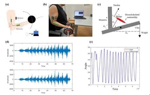
Fig. 1. (a) schematic diagram of sensor configuration and data acquisition scheme; (b) field experimental photo; (c) musculoskeletal model of elbow flexion; (d) collected EMG of biceps brachii in test; (e) collected elbow joint angle in test.
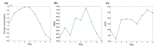
Fig. 2. (a) RMS of the energy signal of randomly selected subject varies with the reps of curling; (b) RMS of torque signals of randomly selected subjects with reps of curling; (c) ZCR of flexion torque of randomly selected subject with reps of curling.
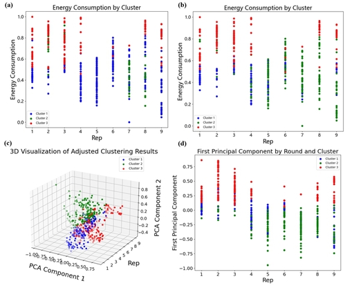
Fig. 3. (a) Clustering results using original nine features. (b) Clustering results of the eight-dimensional features, with Slope of torque excluded. (c) 3D visualization of well-distinguished fatigue clustering. (d) Visualization of fatigue clustering after dimensional reduction based on PCA results.
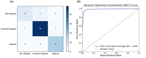
Fig. 4. (a) Confusion Matrix; (b) ROC curve of fatigue prediction.
This result indicates that the biceps brachii torque and power model, constructed using only angle sensors, has enabled effective identification and prediction of muscle fatigue, yielding promising results. Compared to traditional methods with EMG or sEMG signals, the collection and processing of angle signals proved more convenient, particularly suitable for field training environments. It can be consequently concluded that, biceps brachii torque and power model offers a complementary approach in the study of skeletal muscle fatigue and provides valuable insights for research in sports rehabilitation, which may pave the way for novel perspectives and applications in muscle fatigue recognition in future research.
Paper link:https://doi.org/10.1016/j.wees.2024.12.005